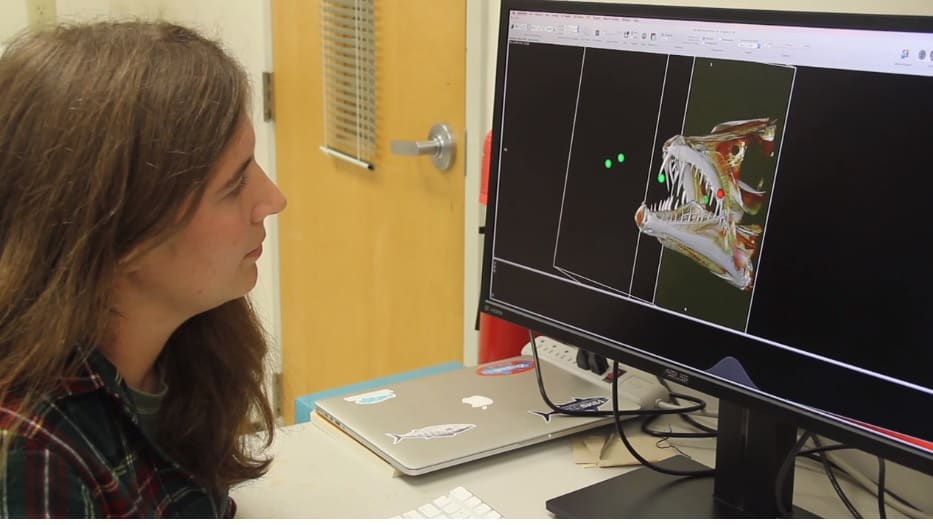
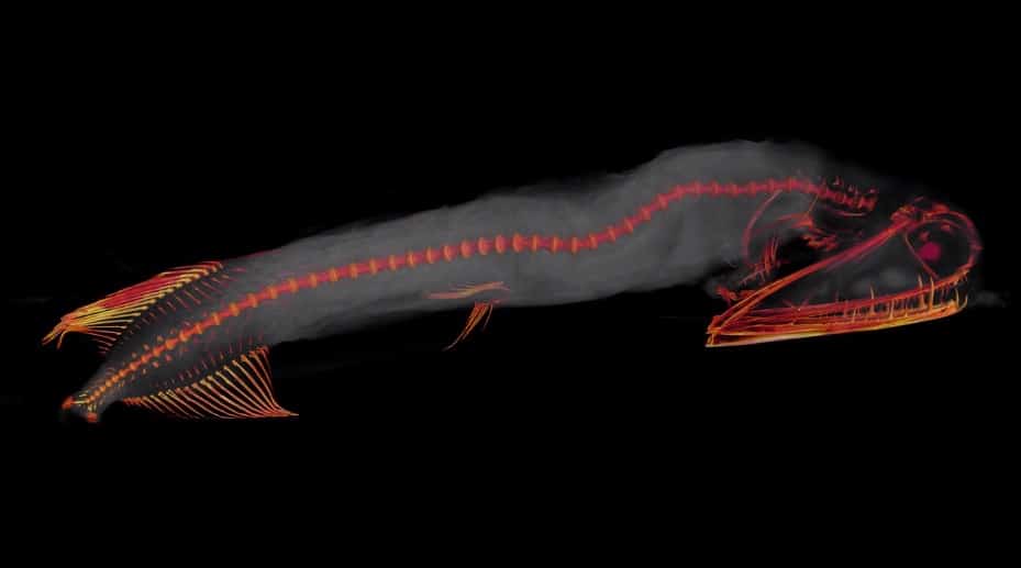
The College of William and Mary’s Virginia Institute of Marine Science (VIMS) is using 3D computer models to “digitally dissect” tiny, larval fish. But they’re calling on crowdfunding to bring that technology to researchers and schoolchildren in the Chesapeake Bay region.
A Look Inside will use micro CT scanning to visualize internal structures like muscles, bones and nerves. Scans can visualize a fish’s stomach contents, and indicate the number and size of eggs in female fish. This gives researchers brand new information about these tiny fish.
With $10,000 of funding, William & Mary undergraduate students will be able to CT scan hundreds of larval and juvenile fish from VIMS at the University of Florida’s state-of-the-art CT scanning facility. Students can then develop 3D computer models that can be used by students and researchers to study the fish up close.

An additional $5,000 in funding will allow the project to print larger-than-life handheld 3D models of the fish, to be used at William & Mary, as well as high schools in James City, York and Gloucester Counties in Virginia.
If you want to contribute to A Look Inside’s $15,000 goal, or see more images of the digitally-scanned fish, click here.
-Meg Walburn Viviano


